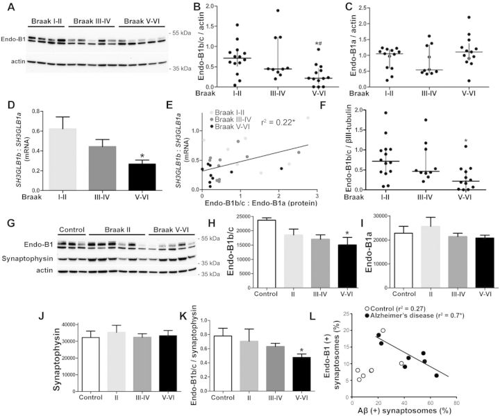
Figure 7.
Expression of neuron-specific endophilin-B1 isoforms is decreased in Alzheimer’s disease. (A) Representative western blot analysing endophilin-B1 protein expression in the inferior parietal lobule cerebral cortex of patients at various Braak stages. Actin was used as a loading control. (B and C) Scatter plot with median and interquartile range of endophilin-B1b/c (B) and endophilin-B1a (C) protein expression. One-way ANOVA showed significant difference in endophilin-B1b/c due to disease progression. *P < 0.05 versus Braak I-II; #P < 0.05 versus Braak III–IV, one-way ANOVA with Tukey post hoc test (n = 10–14 patients per group). (D) The ratio of SH3GLB1b to SH3GLB1a mRNA is decreased in patients with Alzheimer’s disease. *P < 0.05 versus Braak I–II, one-way ANOVA with Tukey post hoc test (n = 7–9). (E) Endophilin-B1b/c to endophilin-B1a protein ratio correlates with SH3GLB1b to SH3GLB1a mRNA ratio. *P < 0.05, Pearson correlation (n = 25). (F) Scatter plot with median and interquartile range of endophilin-B1b/c protein expression normalized to the neuronal cytoskeletal marker βIII-tubulin. One-way ANOVA showed significant difference in endophilin-B1b/c due to disease progression. *P < 0.05 versus Braak I–II. (G) Enriched synaptosome preparations from cortices of control, Braak II, Braak III–IV (not shown), and Braak V–VI patients were analysed for endophilin-B1 expression by western blot. (H and I) Endophilin-B1b/c (H), but not endophilin-B1a (I) expression was decreased in synaptosomes from Braak V–VI patients. One-way ANOVA showed significant difference in endophilin-B1b/c due to disease progression. *P < 0.05 versus control, one-way ANOVA with Tukey post hoc test (n = 6–8). (J) Synaptophysin was not altered in Alzheimer’s disease patient synaptosomes. (K) Endophilin-B1b/c expression was decreased relative to synaptophysin expression in Braak V–VI patients. *P < 0.05 versus control, one-way ANOVA with Tukey post hoc test (n = 6–8). (L) Synaptosomes from controls and Alzheimer’s disease (Braak V–VI) patients were sorted using flow cytometry with antibodies against amyloid-β and endophilin-B1. There was a negative correlation between the number of endophilin-B1 (+) synapses and amyloid-β (+) synapses in the Alzheimer’s disease group only. *P < 0.05, Pearson correlation (n = 7).

