Figure 5.

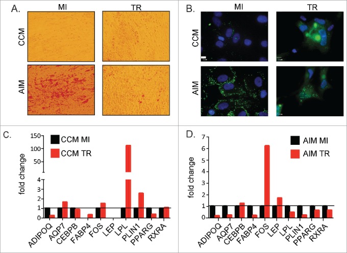
Effects of HCMV infection on adipogensis in ASCs (A) ASCs were cultured in CCM or AIM media for 14 d and mock-infected or infected with HCMV TR strain at MOI = 1. Lipid droplet accumulation was detected using Oil red O staining solution. Images were captures using brightfield settings on an inverted microscope. (B) ASCs were cultured in CCM or AIM media for 14 d and mock-infected or infected with HCMV TR strain at MOI = 1. Lipid droplet accumulation was detected using BODIPY staining solution. Images were captures using a fluorescent inverted microscope. Scale bars = 10 μm. (C) Analysis of ASC mock-infected or infected with TR for 14 d at a MOI = 1. Cellular mRNA was processed for analysis on a custom RT-PCR array designed to examine adipogenic related genes. Fold change of infected cells is relative to mock-infected cells after normalization to house-keeping genes (GAPDH and TBP). Data is from one representative donor out of 3 independent donors. (D) Analysis of ASC mock-infected or infected and then cultured in differentiation media with HCMV TR for 14 d at a MOI = 1. Cellular mRNA was processed for analysis on a custom RT-PCR array designed to examine adipogenic related genes. Fold change of infected cells is relative to mock-infected cells after normalization to housekeeping genes. Data is from one representative donor out of 3 independent donors.
