
Figure 1.
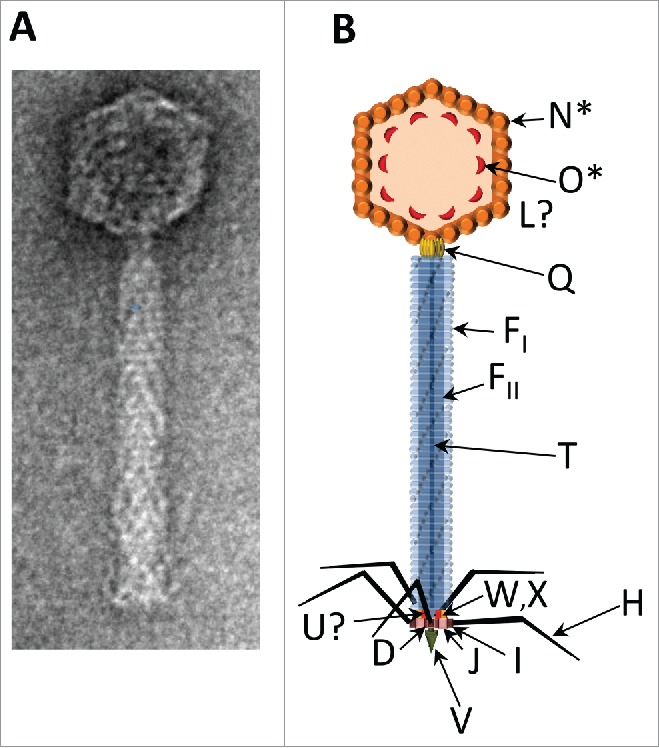
The P2 virion. (A) Electron micrograph of P2, negatively stained with uranyl acetate. This image was generously provided by Dr. Terje Dokland. (B) Schematic illustration of the P2 virion. Arrows indicate the known locations of virion proteins. GpN* is the cleaved form of gpN that constitutes the major capsid protein; gpO* is the fragment of scaffold that remains in the capsid after cleavage. The location in the capsid of the head completion protein gpL is unknown. The dodecameric connector or portal protein, gpQ, lies at the head/tail junction. The tail tube gpFI and tail sheath gpFII are polymerized around the tape measure protein gpT. Arrangement of baseplate components gpW, gpX, gpI, gpJ, gpU, gpD is based on EM studies, known protein-protein interactions, and the location of homologous proteins in the baseplate of bacteriophage T4; assignment for the location of gpU is still tentative. The tail spike is a trimer of gpV, and gpH makes up the tail fibers.