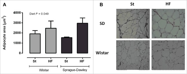
Figure 6.
Mesenteric adipocyte's area (A) of Wistar and Sprague-Dawley (SD) rats fed either with standard (St) or high-fat (HF) diet during 17 weeks. Data are presented as mean ± SEM (n = 6 rats per group). (B) Representative images of hematoxylin and eosin stained-adipose tissue sections for each experimental group.

