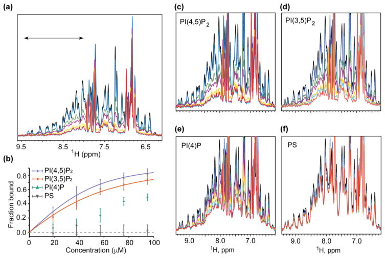
Figure 1.
Influence of lipid constituents on MA binding to liposomes. (a) HIV-1 MA binds with increasing affinity to PC liposomes containing increasing molar percentages of PI(4,5)P2: black, free MA in solution; blue, MA + PC liposomes; green, MA + PC-1%PI(4,5)P2; purple, MA + PC-2%PI(4,5)P2; yellow, MA + PC-3%PI(4,5)P2; cyan, MA + PC-4%PI(4,5)P2; red, MA + PC-5%PI(4,5)P2. (b) The spectral region between 9.5 and 8.0 ppm, corresponding to amide signals of structured residues and indicated in (a), was integrated and used to calculate Kd(apparent) binding affinities. (c–f) 1H NMR spectra of MA free in solution (black) and in the presence of PC liposomes in the absence (blue) and presence of increasing amounts of lipid constituents (1 mol%, green; 2 mol%, purple; 3 mol%, yellow; 4 mol%, cyan; and 5 mol%, red).

