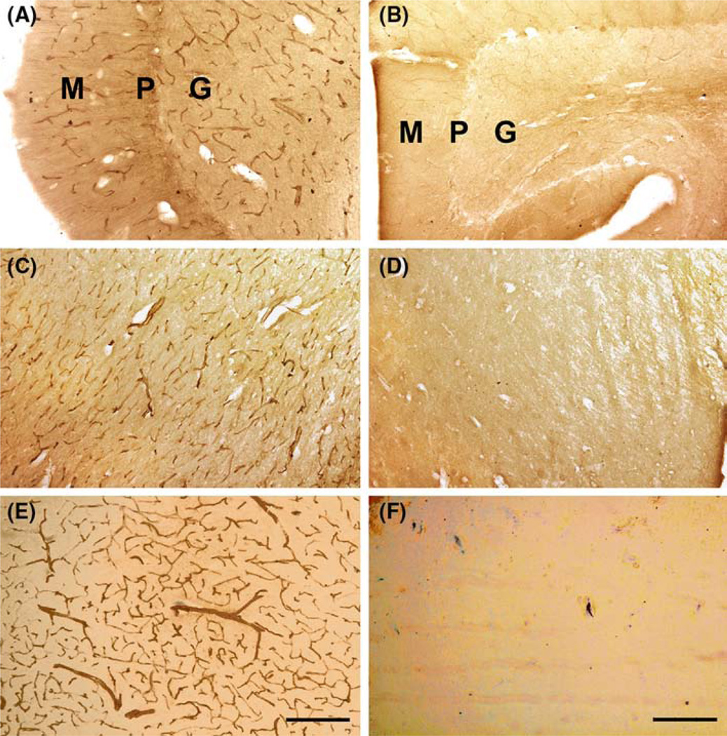
Fig. 4.
Postmortem sections of the cerebellum (HSB-4640 ASD 8.5 year; UMB-1706 control 8.6 year); midbrain/pons and fusiform cortex (UMB4305 ASD 12.9 year; UNB1790 control 13.7 year) from ASD (a, c, e) and control (b, d, f) donors were reacted with nestin antibody. Nestin-positive blood vessels were seen only in the ASD donors in all regions examined. In the cerebellar cortex, the nestin-positive vessels were seen in molecular (M), Purkinje (P) and granular (G) layers in ASD (a) but not control donors (b). In a midbrain/pons section, nestin positive vessels were seen in the midbrain tegmentum in ASD (c) but not in control (d) donors. In fusiform cortex the nestin-positive vessels were seen in layer IV/V from the fusiform cortex in ASD (e) but not in control donors (f). Scale bar 50 µm

