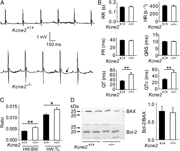
Figure 3.
Kcne2 deletion stimulates baseline cardiac remodelling. (A) Representative surface ECG traces from Kcne2+/+ and Kcne2−/− mice showing prolonged T wave (arrow) in the latter (n = 12–17 each genotype). (B) Actual ECG parameters measured from mice (n = 12–17) showing QT and QTc prolongation in Kcne2−/− mice. **P < 0.01. All other group comparisons P > 0.05. HR, heart rate; PR, PR interval; RR, RR interval. (C) Mean heart-weight to body-weight (HW:BW) and heart-weight to tibia-length (HW:TL) measurements for Kcne2+/+ and Kcne2−/− mice (n = 13–17). *P < 0.05 between genotypes; **P < 0.01 between genotypes. (D) Left: representative western blots showing baseline ventricular BAX and Bcl-2 expression, one mouse per lane. Right: mean band density from blots as in left, n = 6 per genotype.

