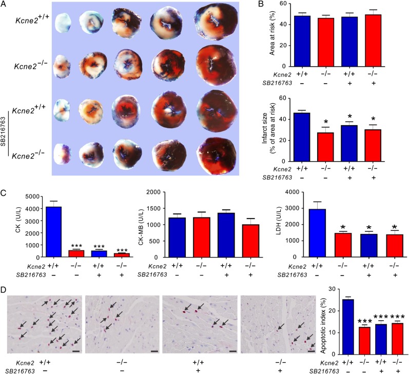
Figure 5.
The effect of pharmacological inhibition of GSK-3β upon reperfusion in Kcne2+/+ and Kcne2−/− mice. (A) Representative TTC-stained sections isolated from Kcne2+/+ and Kcne2−/− mouse hearts subjected to 45 min of LAD ligation followed by 180 min of reperfusion in the presence (+) or absence (−) of SB216763. (B) Upper: AAR expressed as a percentage of LV area. Lower: quantification of myocardial infarct size expressed as a percentage of LV AAR (n = 5–7, each group). All data were expressed as mean ± SEM. *P < 0.05 compared with Kcne2+/+ mice (by one-way ANOVA). (C) Post-IRI mean serum levels of CK, CK-MB, and LDH of Kcne2+/+ and Kcne2−/− mice with (n = 5–6) or without SB216763; values for Kcne2+/+ and Kcne2−/− mice without inhibitors are repeated from Figure 1, for comparison. *P < 0.05, ***P < 0.001 compared with Kcne2+/+ mice (by one-way ANOVA). (D) Left: representative TUNEL-stained heart sections of mice in the presence (+) or absence (−) of SB216763 post-surgery. Arrows indicate TUNEL-positive cardiomyocytes (red). Scale bars, 10 µm. Right: graph showing the averaged percentage of TUNEL-positive cells in the ischaemic regions of LVs. Each group: n = 4–5. ***P < 0.001 compared with Kcne2+/+ mice (by one-way ANOVA). Values for Kcne2+/+ and Kcne2−/− hearts before SB216763 administration are repeated from Figure 1H for comparison.

