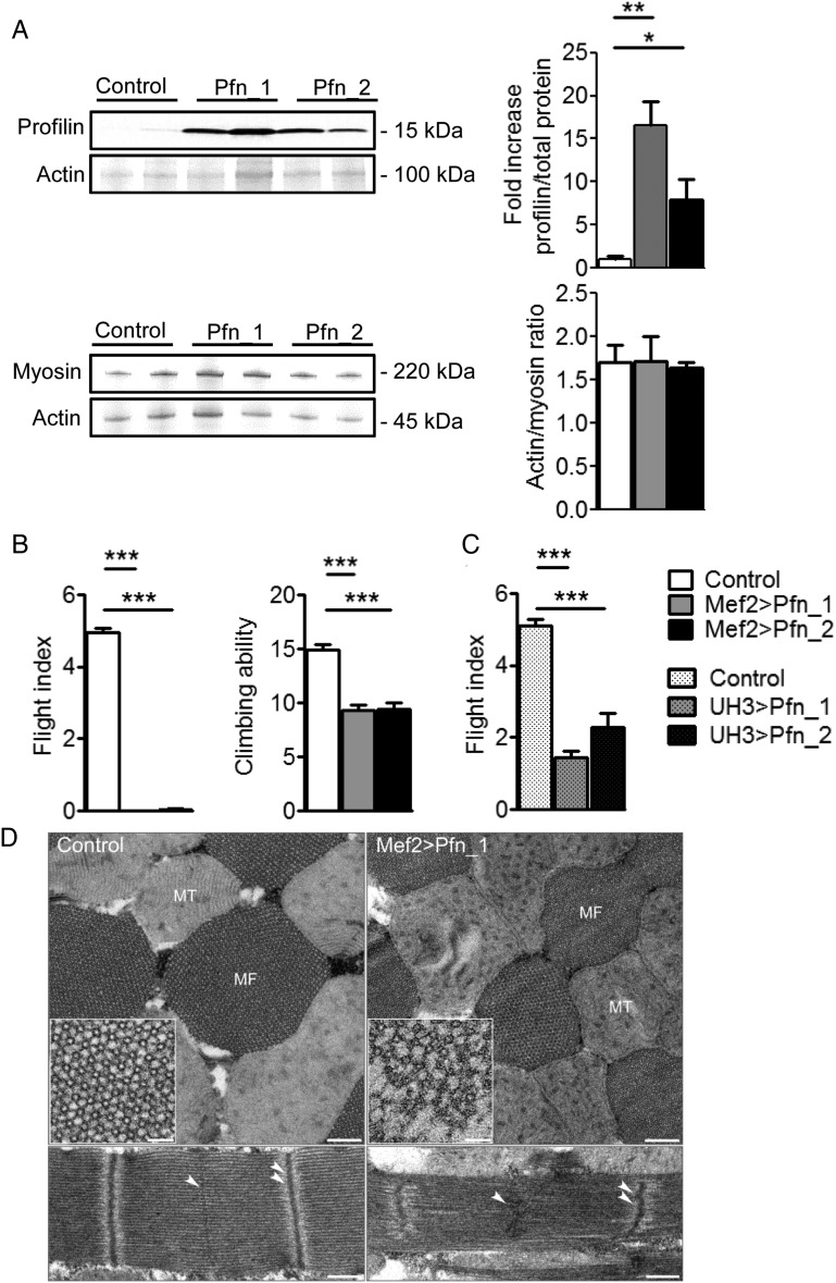
Figure 3.
Overexpression of profilin in Drosophila IFM impairs muscle function and ultrastructure. (A) Western blot analysis showed increased profilin in whole Mef2 > Pfn_1 and Mef2 > Pfn_2 transgenic flies (top) (n = 5, *P < 0.05, **P < 0.01; one-way ANOVA with the Bonferroni post hoc test). Actin/myosin heavy chain ratios remained unchanged in flies with muscle-restricted profilin overexpression compared with control (bottom) (n = 5). (B) Two-day-old Mef2 > Pfn_1 and Mef2 > Pfn_2 flies were unable to fly and demonstrated significantly reduced climbing ability (n = 35–64, ***P < 0.001; Kruskal–Wallis test with Dunn's post hoc test). (C) UH3-GAL4-mediated overexpression of profilin significantly diminished flight ability (n = 54–83, ***P < 0.001; one-way ANOVA with the Bonferroni post hoc test). (D) Representative electron micrographs of transverse sections of Mef2 > Pfn_1 IFMs (top) show that the double hexagonal lattice of myofilament arrangement was less ordered and thin and thick filaments were missing on the outer edges of the myofibril (inset) relative to control. Moreover, there was Z-band buckling and M-line distortion in longitudinal sections (bottom). Single arrowheads delineate an M-line and double arrowheads a Z-line. MT, mitochondrion; MF, myofibril. Scale bars, 500 nm and 250 nm for longitudinal and transverse sections, respectively, and 50 nm in the inset.

