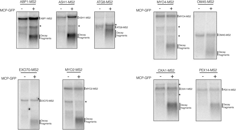
FIGURE 1.
Decay fragments are detectable in mTAG mRNAs when the MCP-GFP plasmid is present. Total RNA was isolated from yeast strains harboring MS2-tagged mRNAs and visualized by Northern blotting using a 1.5% formaldehyde agarose gel. Right lane of each blot is total RNA isolated from strains transformed with pUG36-CP-GFP×3, while the left lane is obtained from untransformed strains. The mRNAs analyzed on the same blot are grouped together. Visualization of the MS2-tagged mRNAs and decay products was done using the 12×MS2 probe (5′-CTGCAGACATGGGTGATCCTCATGTTTTCT-3′). Strains were grown to OD600 0.6 to 0.8 in minimal media (−MCP) or –ura minimal media (+MCP) at 30°C. (*) Indicates background bands.

