FIGURE 3.
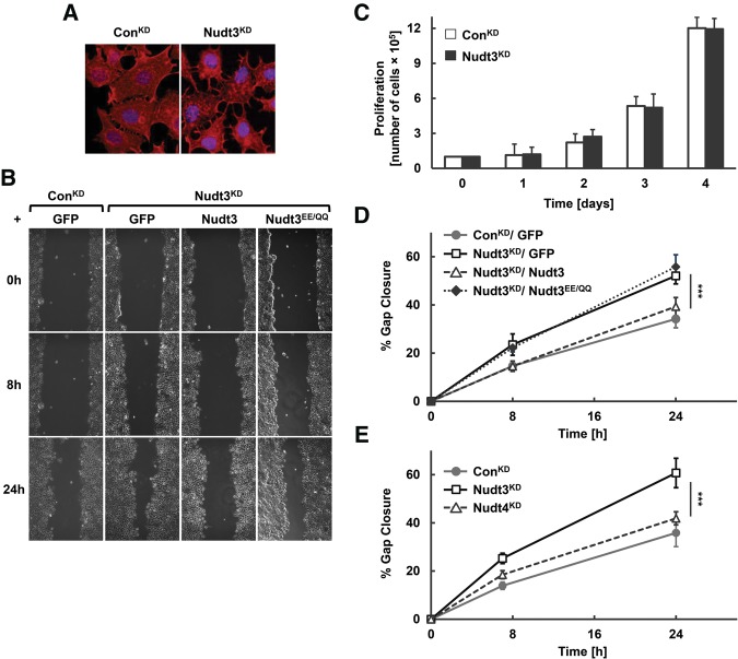
Nudt3 is involved in reducing MCF-7 cell migration. (A) Immunofluorescence of filamentous (F)-actin (red) and DNA (blue) in proliferating ConKD and Nudt3KD MCF-7 cells. (B) Motility of ConKD or Nudt3KD MCF-7 cells complemented with the indicated proteins were analyzed by an in vitro scratch-wound healing assay. The cells were cultured to confluent cell monolayers, treated with mitomycin C to inhibit cell division, scratched and photographed at 0, 8, and 24 h after wounding. (C) Proliferation of ConKD and Nudt3KD MCF-7 cells was quantified by counting the number of cells at day 1, 2, 3, and 4 after initial seeding of 1 × 10⁵ cells per 35-mm dish. Data are representative of three independent experiments. (D,E) The percentage of gap closure for Nudt3KD and Nudt4KD cells were quantitated with ImageJ software. At least five different wounds were quantified for each time point. Data are representative of three independent experiments ±SD. P-values are denoted by asterisks. (***) P < 0.001 (Student's t-test).

