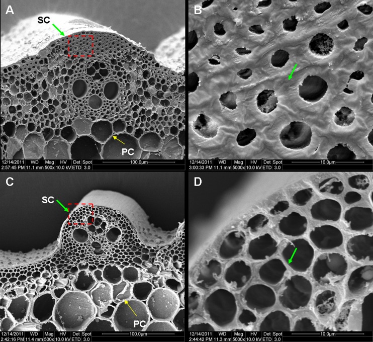
Fig 3. Cross section of a culm viewed under a scanning electron microscope.
(A, B) Cross section of a wild-type culm. (C, D) Cross section of an S1-24 culm. Magnification in the images is 500× (A, C) or 5000 × (B, D). Green and yellow arrows represented thickened sclerenchyma cell (SC) walls and unthicken parenchyma cell (PC) walls.

