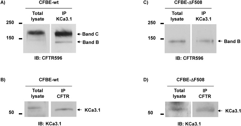
Fig 1. Co-immunoprecipitation of endogenous CFTR and KCa3.1 proteins extracted from CFBE airway cells.
Immunoblots showing CFTR and KCa3.1 proteins extracted from CFBE bronchial cells expressing wt-CFTR (A, B) and F508del-CFTR (C, D). Membranes were blotted with anti-CFTR (mAb 596 from CFFT, 1:1000, A, C) and anti-KCa3.1 (Alomone, 1:300, B, D) antibodies. Endogenous expression of CFTR and KCa3.1 proteins in the CFBE-wt and CFBE-ΔF508 cell lysates are presented in lane “Total Lysate”. Immunoprecipitation of endogenous CFTR using anti-CFTR antibody followed by co-immunoprecipitation of KCa3.1 is illustrated in lane IP CFTR (B, D), while immunoprecipitation of endogenous KCa3.1 (using anti-KCa3.1 antibody) followed by co-immunoprecitation of CFTR is shown in lane IP KCa3.1 (A, C). Note that the same lysate and IP samples were used in the upper and lower parts of the membranes, blotted with CFTR and KCa3.1 antibodies, respectively.

