
Figure 3.
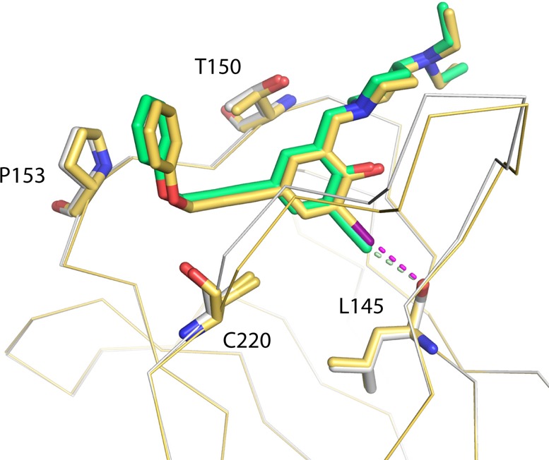
Binding modes of PK5176 (2, yellow carbons) and PK5211 (3, green carbons) in complex with the p53-Y220C DNA-binding domain by X-ray crystallography. Both compounds share a nearly identical binding mode, with 3 shifted rigidly upward away from Leu145 because of the increased length of its terminal ethynyl group. The I···O halogen bond between 2 and the protein is shown as a purple broken line, and the CH···O hydrogen bond between 3 and the protein is shown as a light green broken line.