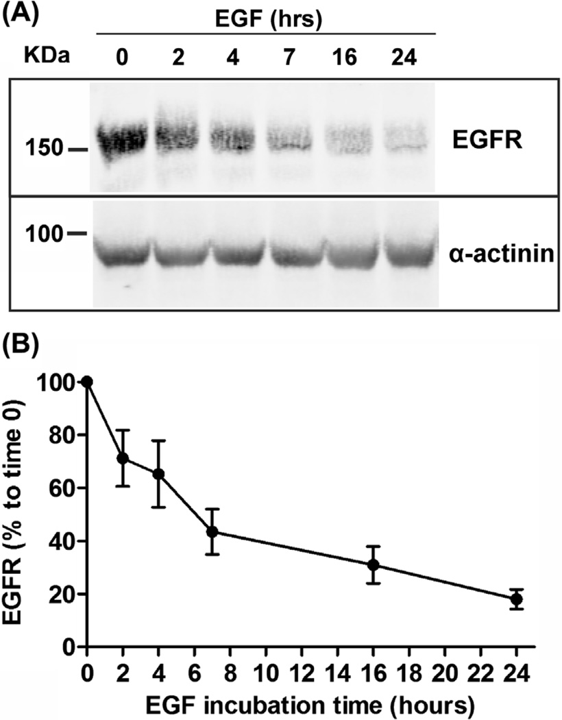
FIGURE 5. Epidermal growth factor (EGF)-induced EGF receptor (EGFR) degradation.
UMSCC2 cells were incubated with 80 ng/mL EGF for 0–24 h. (A) EGFR was detected by western blotting in cell lysates using antibodies 1005. α-actinin is as loading control. (B) Quantification of EGFR immunoreactivity from two experiments including the data presented in (A).

