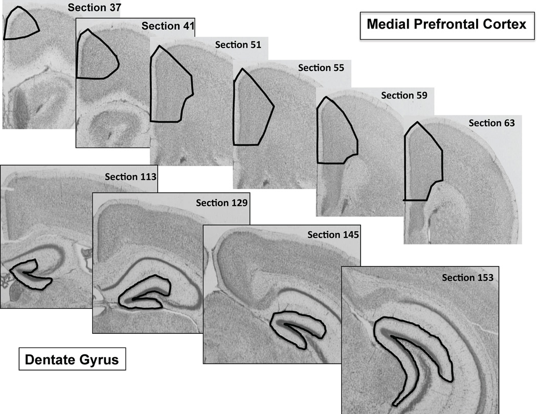
Figure 1.
Coronal sections of a representative mouse brain stained by the Nissl method taken from the medial prefrontal cortex (black contours in the upper row) and dentate gyrus of the hippocampus (black contours in the lower row). The black contours represent the entire rostral-to-caudal extent of these areas in which the total number of neurons and glial cells were counted using the optical fractionator method of StereoInvestigator software.

