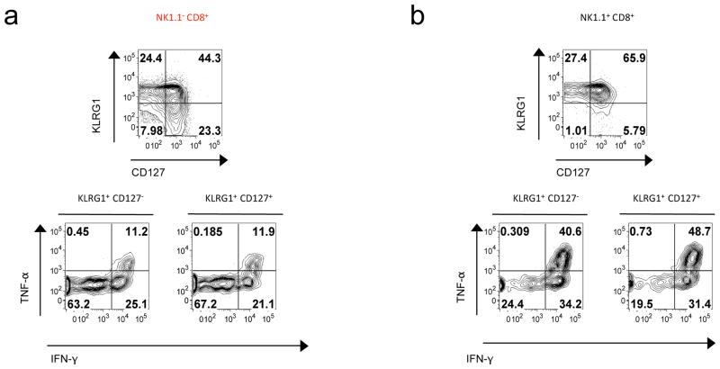
Figure 3. NK1.1+ CD8+ T cells consist of activated and effector cells.
(a–b) B6 mice were infected by LCMV and their spleens analyzed 9 days later by flow cytometry. Expression of surface expression of KLRG1 and CD127 as well as IFN-γ and TNF-α production by H2-Db(NP396–404) tetramer+ CD8+ T cells after 3 hours of culture with LCMV NP396 peptide-pulsed APC. These results are representative of 2 independent experiments with 3mice per group.

