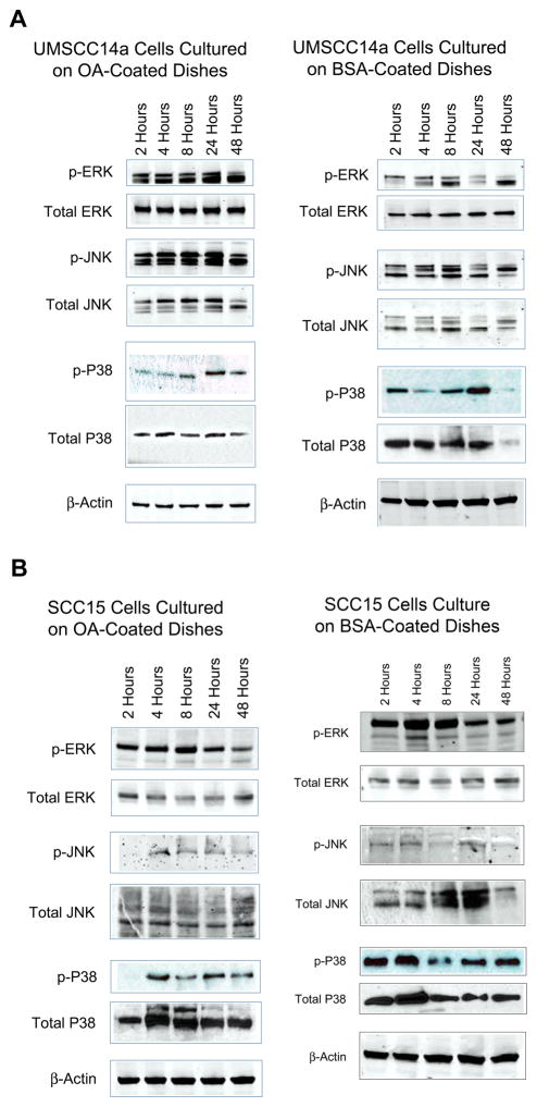
Figure 5. OA activates MAPKs in OSCC.
OSCC cells were allowed to adhere to tissue culture dishes coated with rhOA or BSA. Cells were treated with pervanadate 30 minutes before cellular lysates were harvested after 2, 4, 8, 24 and 48 hours. Lysates were probed with antibodies to MAPKs. (A) As compared to control, UMSCC14a cells adhered to rhOA demonstrated significant activation of ERK and JNK MAPKs that peaked between 8 and 24 hours. (B) As compared to control, SCC15 cells adhered to rhOA demonstrated delayed and less MAPK activation.

