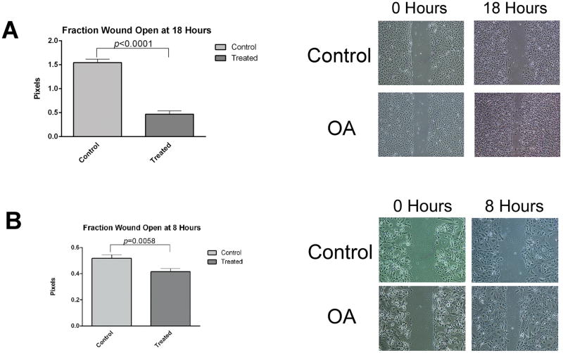
Figure 6. OA promotes migration of OSCC cells.
OSCC cells were plated at confluence on rhOA- or BSA-coated dishes. Wound healing migration assays were used to determine OA’s effect on cell migration. Graphs indicate means ± standard errors. OA accelerated migration of OSCC cells in monolayer wound healing assays. (A) UMSCC14a cells exhibited migration of individual cells as well as groups of cells. Die-back of UMSCC14a accounted for larger wounds at 18 hours in plates coated with BSA. (B) SCC15 cells exhibited primarily group migration.

