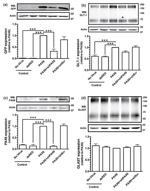
Figure 3. Effects of Pax6 and shRNA against Pax6 on the levels of eGFP or GLT-1 protein.
Cortical astrocytes from BAC GLT-1 eGFP transgenic mice were infected with different combinations of lentiviral vectors, including those that contain: dsRED & empty virus, Pax6 & empty virus, Pax6 and shRNA directed against Pax6, or Pax6 and a scrambled shRNA (shScr). Cells were harvested after 10 d and the levels of eGFP (a) or GLT-1 (b) were analyzed by Western blot. Fifteen μg of cell lysate was loaded to each lane. Top, Representative Western blots for eGFP or GLT-1. Bottom, Summary of eGFP or GLT-1 protein levels normalized to actin and expressed relative to the levels observed in astrocytes infected with Pax6. Cell lysates were also obtained from astrocytes that were not transduced with lentivirus and used as an additional control (No Virus). Pax6 (c) and GLAST (d) protein levels were also analyzed in these same specimens. Top, A representative Western Blot; bottom, graphical summary of data. Fifty μg of cell lysate was loaded in each lane for Western blot analysis of Pax6. No other immunoreactive bands were observed in these blots. Data are the mean ± SEM of three independent experiments. ***p < 0.001 compared to corresponding Pax6 infected astrocytes.

