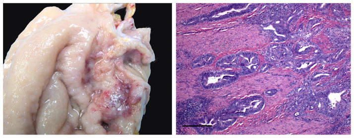
Figure 1.
Animal 9 gastric adenocarcinoma. Left panel indicates gross image of stomach from animal #9 depicting an ulcerated raised mass. Notice multifocal areas of hemorrhage and necrosis. Right panel depicts photomicrograph of a section of gastric mucosa and submucosa. The neoplastic epithelial cells extend deep into the submucosa and muscular layer (bar = 100 microns).

