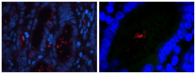
Figure 2.
FISH for Helicobacter genus of gastric tissue from study animals. Photomicrographs of positive hybridization of Helicobacter genus level probes to gastric tissue. Left and right panels are from animals #10 and #9, respectively. The left panel is at 630× magnification and the right panel is at 1000× magnification. Both hybridized probes demonstrate tightly coiled morphology typical of Helicobacter suis. (Red = FISH probe hybridization, Blue = DAPI, Green = Background).

