
Figure 3.
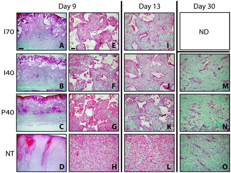
Representative images of trichrome staining at day 9 with paired magnifications of 2× (left) and 20× (right). Wounds at days 13 and 30 are shown at 20× with the exception of Section M, which shows a day 30 wound injected with a scaffold with 40% sucrose at a 2× magnification. Mature collagen is intense green, nascent collagen is pale green, cytoplasm and fibrin are red or pink. PUR scaffold remnants remain unstained and appear white with angular profiles. Scale bar in A (2× magnification) = 1000 μm; Scale bar in E (20× magnification) = 100 μm.