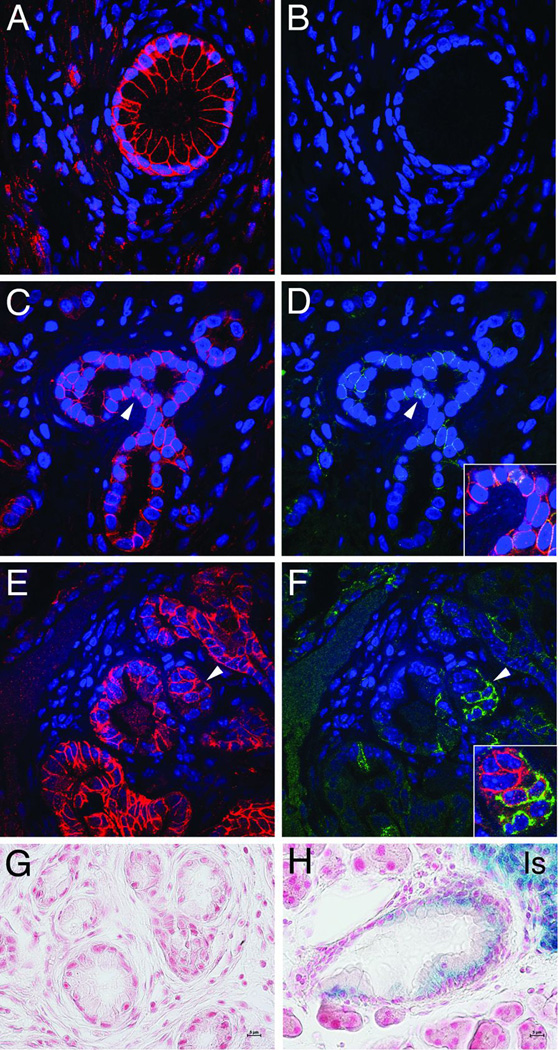
Figure 1. E- and N-cadherin expression in human and murine PanIN.
Double immunofluorescence analysis of E-cadherin (antibody from Santa Cruz) and N-cadherin (antibody from Invitrogen) in human (A–D) and murine (E, F) pancreas specimens. Pancreas samples were obtained from patients at Johns Hopkins Medical Center (JHMC) following approval by JHMC IRB. E-cadherin was present both in normal pancreatic duct (A) and PanIN (C). By contrast, N-cadherin was absent from normal ducts (B), but found upregulated in a subset of PanIN lesions (D). Arrowheads point to areas shown in merged image (insets). E-cadherin (E) and N-cadherin (F) expression in mPanIN from K-rasG12D; Pdx1/Cre (KC) mice. Note ductal cells with decreased E-cadherin and increased N-cadherin. Histological analysis of β-galactosidase-stained mPanIN from Ncad+/+ (G) and NcadlacZ/+ (H) KC mice. N-cadherin LacZ reporter showed a positive signal (blue) in ductal cells and islets of Langerhans (Is) in the KC NcadlacZ/+ mice (H) whereas KC mice lacking the LacZ reporter served as a negative control (G).

