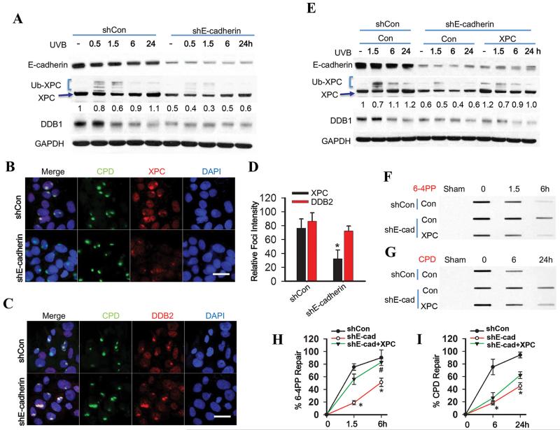
Fig. 2. E-cadherin regulates 6-4PP repair but not CPD repair via increasing XPC availability.
(A) Immunoblot analysis of E-cadherin, XPC, Ub-XPC (polyubiquitinated XPC), DDB1 and GAPDH in HaCaT cells transfected with shCon or shE-cadherin at 0, 0.5, 1.5, 6 and 24 h post-UVB (20 mJ/cm2). The results were obtained from three independent experiments. (B-C) Immunofluorescence assay of the subnuclear colocalization of CPD and XPC (B) and CPD and DDB2 (C) in HaCaT cells transfected with shCon or shE-cadherin at 0.5 h post-UVC (10 mJ/cm2) through a 5 μm micropore filter. Scale bar, 10 μm. (D) The relative intensity of XPC and DDB2 focus was calculated by analyzing 100 foci and normalized to that of CPD (n= 100, bar: SD). (E) Immunoblot analysis of E-cadherin, XPC, DDB1 and GAPDH in HaCaT cells transfected with shCon, shE-cadherin, or the combination of shE-cadherin with XPC plasmids at 0, 1.5, 6 and 24 h post-UVB (20 mJ/cm2). The results were obtained from three independent experiments. XPC protein levels in A and E were quantified using ImageJ software (below each band in arbitrary units). (F-G) Slot blot analysis of 6-4PP (F) and CPD (G) in HaCaT cells transfected with shCon, shE-cadherin, or the combination of shE-cadherin with XPC plasmids at 0, 1.5 and 6 h post-UVB (20 mJ/cm2) for 6-4PP and 0, 6 and 24 h post-UVB (20 mJ/cm2) for CPD. (H and I) Quantification of percentage (%) of 6-4PP repair from F and CPD repair from G. *, P < 0.05, compared with shCon group; #, P < 0.05, compared with shE-cadherin together with shCon groups, Student’s t-test and two-way ANOVA.

