
Figure 7.
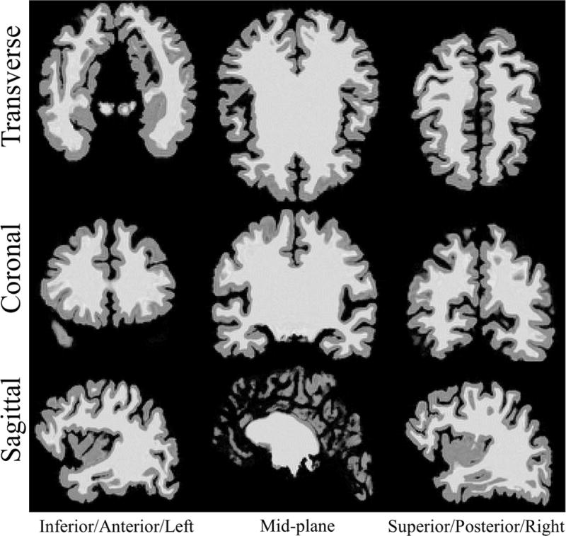
Representative slices of reconstructed volume from 3D MRI simulation using analytical polyhedral brain mesh phantom. Rows top to bottom: Transverse, coronal, sagittal planes. Columns left to right: Inferior, mid-plane, superior slices for the transverse plane. Anterior, mid-plane, posterior slices for coronal plane. Left, mid-plane, right slices for the sagittal plane.