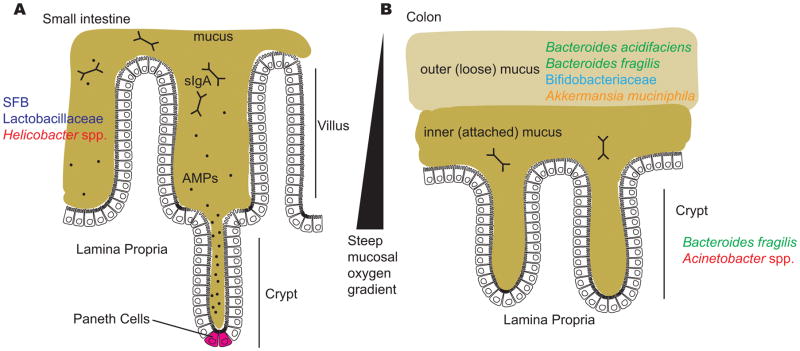
Figure 2. The mucus layers of the small intestine and colon.
Several factors limit the ability of gut bacteria to access host cells, including the mucus layers in the small intestine and the colon; antimicrobial peptides in the small intestine, including those produced by Paneth cells at the base of the crypts; secreted immunoglobulin A (sIgA) in both the small intestine and colon; and a steep oxygen gradient that influences which bacteria are capable of surviving close to the epithelial surface. A) The surface of the small intestine is shaped into villi and crypts and is colonized by certain adherent species, including segmented filamentous bacteria (SFB), Lactobacillaceae and Helicobacter spp. B) The colon has two distinct mucus structures: the outer layer is colonized by mucin-degrading bacteria and is characterized by the presence of Bacteroides acidifaciens, Bacteroides fragilis, Bifidobacteriaceae and Akkermansia muciniphila and the inner layer and crypts are penetrated at low density by a more restricted community that includes Bacteroides fragilis and Acinetobacter spp.

