Fig. 3.
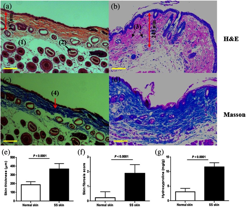
Histopathological features of BLM-induced skin fibrosis. Representative H&E stained skin sections from (a) PBS-injected control group specimen and (b) BLM-injected skin sample, at magnification. Compared to the PBS control group in (a), BLM-injected mice showed increased skin thickness, loss of subcutaneous fat tissue, as well as increased cellular infiltration. Representative sections stained with Masson’s trichrome at magnification from a (c) PBS-injected control group skin specimen and (d) BLM-treated sample. Thickened collagen bundles were observed in the BLM-injected mice, compared to the PBS-injected control mice. The scale bars represent the spatial distance of . The main features of the skin have been shown in (a) to (d), including fat tissue (1), hair follicles (2), inflammatory cells (3), and collagen (4). The skin thickness measurement is also shown in (a) and (b) by the double-headed red arrow; (e) skin thickness, (f) skin fibrosis score, and (g) skin hydroxyproline content were significantly higher in the BLM-SSc mice than in the control mice. Shown data are representative of similar findings from 18 mice from the control group and 13 mice from the BLM-SSc group.

