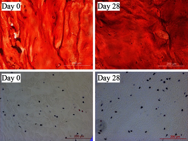
Figure 4.
Extracellular matrix and viability staining after long-term culture. Safranin-O/Fast green and LDH staining on day 0 (left) and day 28 (right). Proteoglycans are stained red, collagen green and cell nuclei black in the upper images. Living cells are stained black and their nucleic acids red in the lower images. Scale bars are 200 μm.

