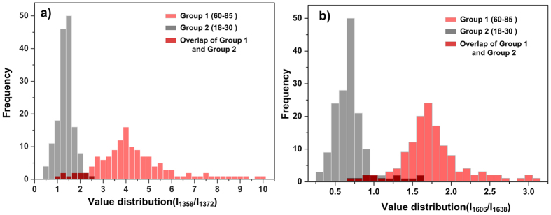
Figure 6. Detection of the oxygenation of haemoglobin using SERS spectroscopy.
(a) Distribution of value of I1358/I1372 for both groups. (b) Distribution of value of I1606/I1638 for both groups. The light red histogram represented Group 1while the grey histogram represented Group 2. The dark red part is the overlap of both groups. The SERS spectra number brought into statistics of Group 1 is n = 138, and that of Group 2 is n = 156.

