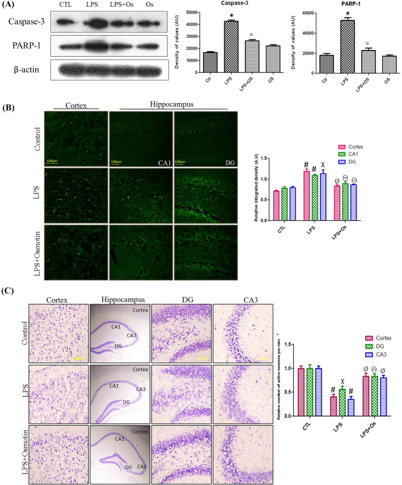
Figure 7.
Osmotin attenuated LPS-induced apoptotic neurodegeneration in the hippocampus of adult mice (A) Shown are representative Western blots probed with caspase-3 and PARP-1 antibodies in the hippocampus of adult mice. The density values are expressed in arbitrary units as the mean ± SEM for the indicated proteins (n = 5 animals per group). Shown are representative photomicrographs of (B) FJB staining (magnification 40× objective field, scale bar = 100 μm) and (C) Nissl staining (magnification 20× objective field, scale bar = 200 μm) for dead and damaged neurons. Images are representative of staining obtained in sections prepared from at least 5 animals per group. Symbols for treatment groups and level of significance are mentioned in the data analysis section of the Materials and Methods.

