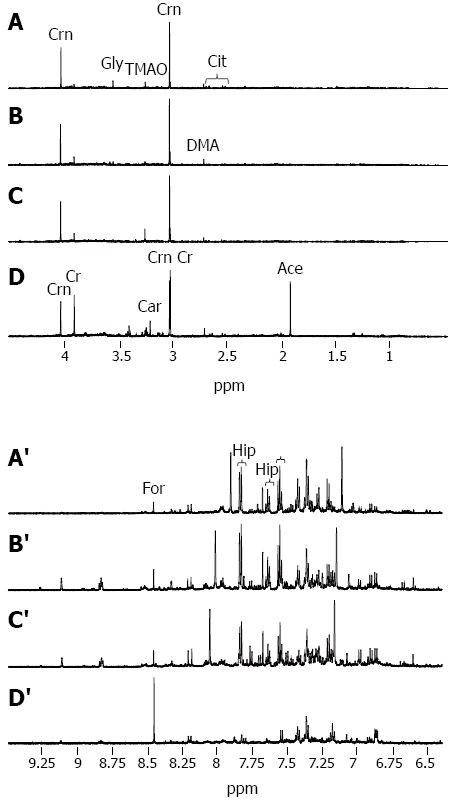
Figure 1.

Illustrative urinary proton nuclear magnetic resonance spectra. From a 35 year old healthy control (CTR) (A, A’); a 22 year old male with chronic hepatitis-B related liver disease (CHB) (B, B’); a 36 year old male with hepatitis B virus (HBV)-cirrhosis (C, C’); and a 50 year old male with HBV-hepatocellular carcinoma (AFP > 30000 mg/dL) displaying (A-D) the aliphatic region 0.5-4.5 ppm and (A’-D’) the aromatic region 6.4-9.5 ppm (D, D’). Each NMR spectrum is scaled independently. The more prominent peaks are assigned and include acetate (Ace), carnitine (Car), citrate (Cit), creatine (Cr), creatinine (Crn), dimethylamine (DMA), formate (For), glycine (Gly), hippurate (Hip), histidine (His), and trimethylamine-N-oxide (TMAO).
