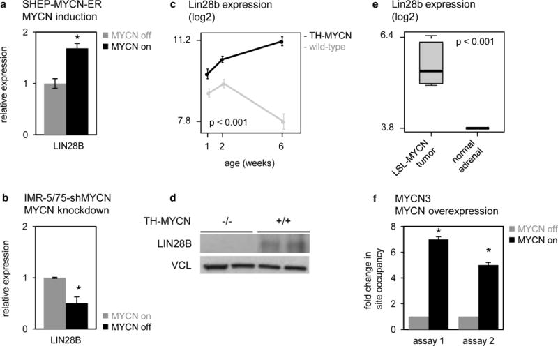
Figure 6. MYCN induces LIN28B expression in neuroblastoma cells.

(a) Relative expression of LIN28B in SHEP-MYCN-ER cells before (gray bars) and after (black bars) induction of MYCN activity. Data represent average fold change of three independent biological replicates +/− standard deviation. T-test, p = 4.2 × 10⁻³. (b) Relative expression of LIN28B in IMR-5/75-shMYCN cells before (gray bars) and after (black bars) silencing of MYCN. Data are presented as mean ± standard deviation of three replicate experiments T-test, p = 5.7 × 10⁻³. (c) Lin28b expression (log2) in sympathetic ganglia containing hyperplastic lesions, and advanced tumors from TH-MYCN+/+ mice at, respectively, 1 and 2 weeks and 6 weeks of age (black), and in normal sympathetic ganglia from wild-type mice at 1, 2 and 6 weeks of age (gray). Data are presented as mean ± standard deviation of four samples. Two-way ANOVA, interaction p = 1.9 × 10⁻⁸. (d) Lin28b protein levels in advanced tumors from TH- MYCN+/+ mice at 6 weeks of age (indicated as “+/+”), and in normal sympathetic ganglia from wild-type mice at 6 weeks of age (indicated as “−/−”). (e) MYCN ChIP was performed on MYCN3 cell line either doxycycline treated (MYCN high) or untreated (MYCN low). Two assays (Supplementary Table 5) for MYCN binding site at LIN28B promoter was designed by analyzing MYCN ChIP-Sequencing database. Negative control IgG ChIP and Input material was also analyzed and used for normalizing the MYCN ChIP data. Two sets of control primers were used to confirm the specificity of immunoprecipitated genomic DNA. Fold enrichment in site occupancy was calculated by comparing normalized MYCN ChIP Cq values between treatments. ChIP-qPCR reactions were performed in triplicates and represented here as mean ± SEM. T-test, p < 0.001.
