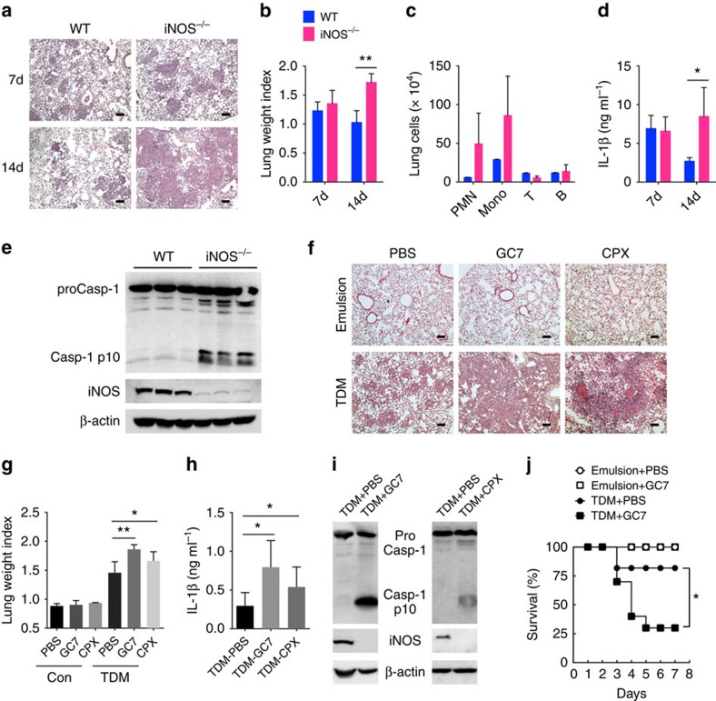
Figure 7. iNOS deficiency and inhibition of eIF5A hypusination aggravate granuloma formation by TDM.
(a–e) WT and iNOS−/− mice were injected intravenously with an oil-in-water emulsion of TDM (n>6 mice/group). (a) Histology of lungs at 7 and 14 days after injection (scale bars, 100μm). (b) TDM-induced lung swelling. On days 7 and 14 after injection of TDM, lung swelling was evaluated by LWI. (c) Identification of leucocyte subsets in lung granulomas on day 14 after TDM injection by flow cytometry. The number of neutrophils (PMN, CD11b+ Ly6G+), monocytes (Mono, CD11b+ Ly6G−), T cells (CD3+) and B cells (CD19+) are indicated. (d) ELISA of IL-1β in lung lysates on day 7 or 14 after TDM injection. (e) Immunoblot analysis of active caspase-1 (Casp1 p10) and iNOS in lysates of lungs from WT and iNOS−/− mice injected with TDM; β-actin serves as a loading control (each lane represents an individual mouse). (f–j) Mice were administered with PBS, GC7 or CPX via intraperitoneal injection, and with oil-in-water emulsion of TDM or emulsion alone, via intravenous injection. On day 7 after the indicated injection, lungs were harvested and experiments were performed. (f) Lung histology was examined by H&E staining (scale bars, 100 μm.) (g) TDM-induced lung swelling. At day 7 after the indicated injection, lung swelling was evaluated by LWI. (h) ELISA of IL-1β in lung lysates on day 7 TDM injection. (i) Immunoblot analysis of active caspase-1 (Casp1 p10) and iNOS in lysates of lungs from mice injected with TDM with/without GC7 or CPX, assessed on day 7 after injection; β-actin serves as a loading control (each lane represents an individual mouse). (j) Lethal systemic inflammation by TDM (n=five mice/group, P-value calculated by Mantel–Cox test). *P<0.05, **P<0.01 (two-tailed unpaired Student's t-test). Data are representative of two experiments (b–d,g–i: mean and s.d. of three to five mice per group).

