Fig. 1.

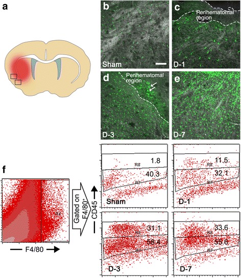
CX3CR1+ cells increase in the peri-hematoma region after ICH. a CX3CR1+ cells were detected in peri-hematoma regions of hemorrhagic brain sections. b–e Representative images on brain sections were obtained from Cx3cr1 +/GFP mice at 1, 3, and 7 days after ICH or sham operation. Three days after ICH, amoeboid cells were detected in the peri-hematoma region (arrows) (Scale bar, 100 μm). f Myeloid cells (F4/80+) in the lesion were analyzed and further divided into CD45-low microglia and CD45-high macrophage populations according to the expression level of CD45 according to flow cytometry. The flow cytometry data are representative of three independent experiments
