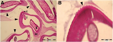
Fig. 1.

Histological section of subcutaneous formation extracted from hypogastrium (PAS staining; Case 1). a Section of the nodule containing the coiled worm (arrowheads) with visible genital tube (g), intestine (i), coelomyar musculature (m) and striated cuticle (c); 100 x. b Detailed view on longitudinal and transversal striation of the cuticle (arrowhead); 400 x
