Fig. 3.

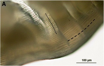
The worm extracted from the nodule on the left middle-finger of the patient (Case 4). a. Detail on cuticular structure with Dirofilaria-specific longitudinal ridges (-----) and transversal striation (∙∙∙∙∙); Photo P. Kotíková

The worm extracted from the nodule on the left middle-finger of the patient (Case 4). a. Detail on cuticular structure with Dirofilaria-specific longitudinal ridges (-----) and transversal striation (∙∙∙∙∙); Photo P. Kotíková