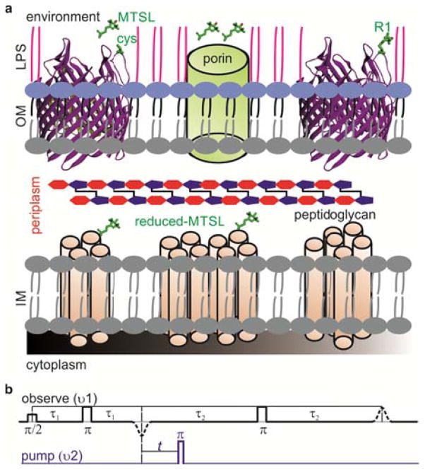
Figure 1.
(a) Structure of the the Gram-negative bacterial cell wall. The outer membrane (OM) is assymetric consisting of an inner phospholopid layer and an outer lipopolysacharide (LPS) layer. The inner membrane (IM) consists of a phosopholipid bilayer containing numerouns α-helical proteins. Exposed cysteines on β-barrel proteins can be labeled by additon of MTSL from outside. Those MTSL molecules which enter periplasm through the porins are reduced. (b) The PELDOR (4-pulse DEER)9 pulse sequence consists of a refocused echo at the observer frequency (black) and its intensity modulation is monitored as a function of the timing of an inversion pulse at the pump frequency (blue).

