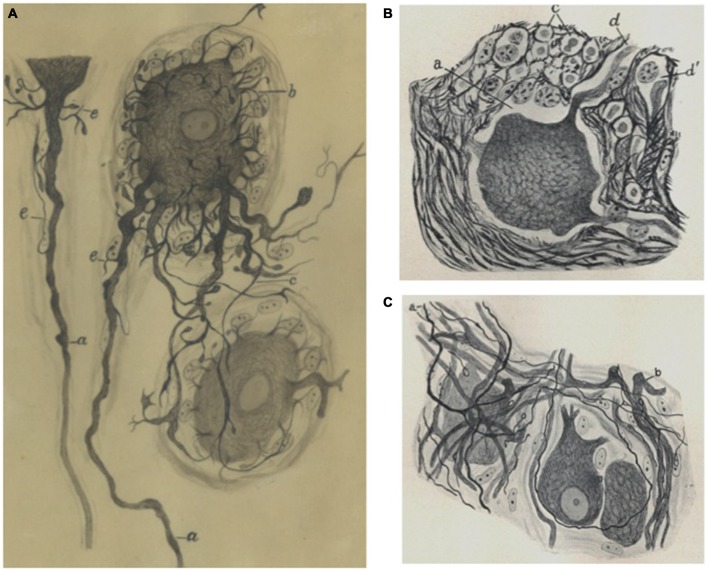
Figure 3.
Sympathetic neurons by Fernando de Castro. (A) de Castro’s hand-made schematic illustration of sympathetic neurons stained with the Cajal’s method, showing short long (a –the axón arises from this dendrite at a distance from the soma, c), short dendrites (b). This image was published in de Castro (1933). (B) Partial view of a sympathetic ganglion (normal condition) of an adult cow (de Castro, 1937). (C) Portion of a sympathetic ganglion with regenerated preganglionic fibers (a) after a vagus-sympathetic crossed anastomosis (de Castro, 1937; A) is part of Archive Fernando de Castro.

