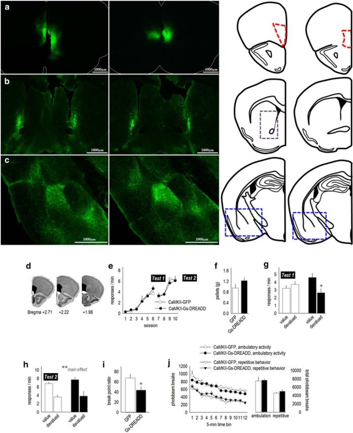
Figure 1.

Chemogenetic stimulation of the mOFC enhances behavioral sensitivity to reinforcer value and PR response requirements. a, Viral vectors expressing GFP were infused into the mOFC, annotated by the anatomical boundaries outlined at right. b, Fluorescing axons were detectable in a stereotyped pattern hugging the medial wall of the dorsal striatum, particularly in the rostral portion highlighted by the gray dashed lines (cf. Schilman et al., 2008). c, Terminals were also detected in the medial compartment of the basal amygdala. The corresponding coronal sections are shown at right. Blue boxes outline the areas shown in the photomicrographs. d, Infection spread for viral vector experiments is represented on images from the Mouse Brain Library (Rosen et al., 2000), with black indicating the largest spread and white the smallest. e, Mice expressing CaMKII-driven AAV–GFP or AAV–Gs–DREADD–mCitrine acquired the instrumental response. Breaks in the response acquisition curves indicate tests for behavioral sensitivity to reinforcer devaluation. f, Mice were fed the reinforcer pellets or regular chow ad libitum before probe tests (devalued and value conditions). Groups did not differ in food consumption. g, Activation of Gs–DREADDs augmented behavioral sensitivity to reinforcer devaluation, decreasing response rates. Meanwhile, control mice did not modify their response patterns. h, A second experience with the reinforcer devaluation procedure and injection stress ultimately resulted in response inhibition in both groups (n = 4–6 per group). i, Activating Gs–DREADDs also reduced breakpoints in a PR test (n = 6–10 per group). j, Locomotor activity was not effected by Gs–DREADDs stimulation. Ambulatory and repetitive photobeam breaks are represented in 5 min bins (left) and 1 h total counts (right). Mice in both instrumental conditioning experiments were tested. Bars and symbols represent means ± SEMs. *p < 0.05, **p < 0.001.
