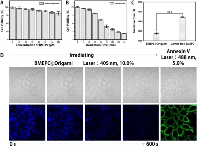
Figure 3.

(A) Cell viability of MCF-7 cells after incubated with different concentrations of BMEPC for 12 h in darkness. (B) Cell viability of MCF-7 cells after irradiated in DMEM (phenol red free) for different durations of time. (C) Time to 50% inhibition of irradiation after incubation with 20 μM carrier-free BMEPC and 1 nM (20 μM BMEPC-loaded) DNA origami complex individually for 12 h, and then irradiated in DMEM (phenol red free), p = 0.0002. (Lamp: 365 nm, 8 W. Optical Density: 0.067 W/cm2. Irradiation distance: 1 cm.) (D) One-photon induced CLSM irradiation (blue) and Annexin V apoptosis fluorescence staining (green) at 405 nm. Adhered MCF-7 cells were incubated with 1 nM (20 μM BMEPC-loaded) DNA origami for 12 h in DMEM, irradiating in DMEM (phenol red free) for 600 s and imaging in Annexin V binding buffer. The scale bars are 25 μm.
