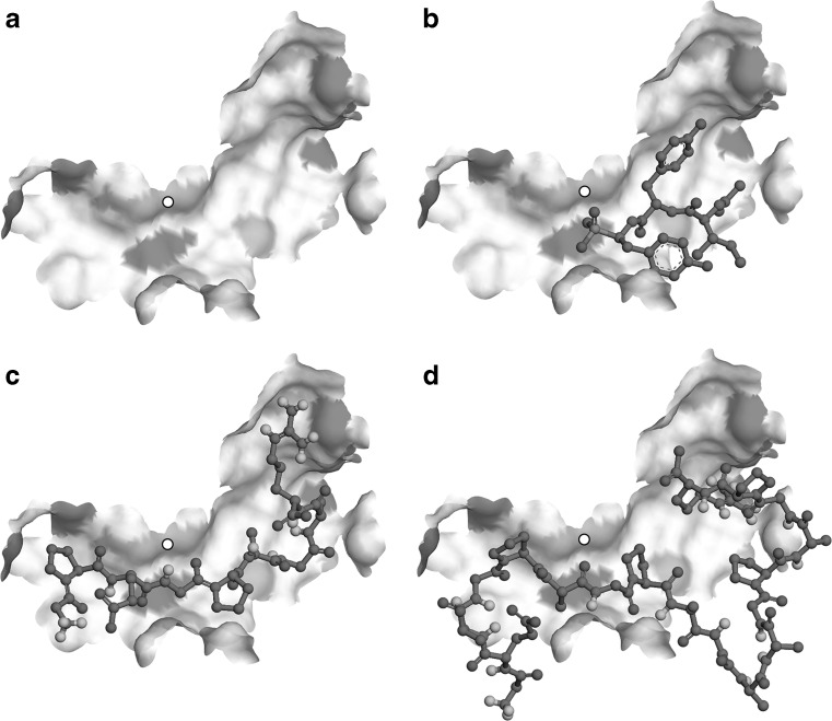
Fig. 2.
Docking poses of ligands onto the active site of ACE. a Apo-enzyme generated by a manual removal of original (K-26) ligand from the co-crystal complex structure is shown in a surface model where ionic residues are highlighted in gray patches. A small white circle indicates the location of Zn2+ co-factor of the enzyme. b K-26 molecule re-docked onto the active site of the enzyme is shown in a ball-and-stick model. Peptide 1 (c) or peptide 2 (d) were also docked into the active site of the same apo-enzyme structure. Light gray atoms are oxygen

