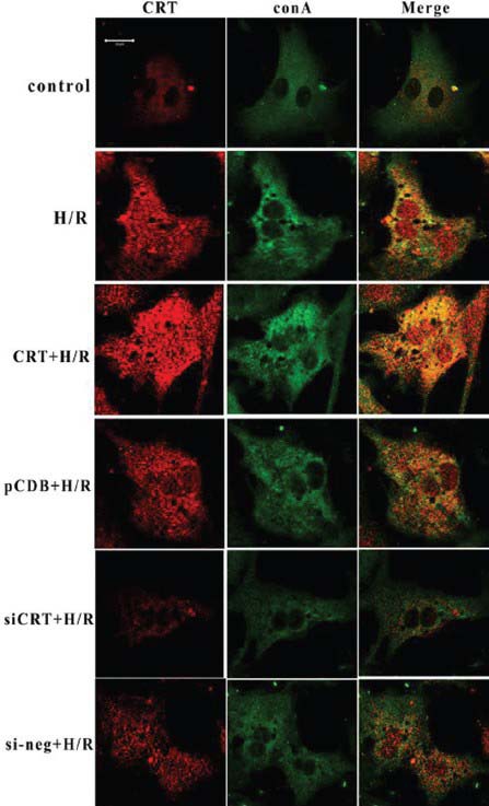
Figure 2.

Co-immunocytoflurescene of concanavalin A (green) and calreticulin (CRT) (Red). During the resting state, the endoplasmic reticulum displayed a homogeneous faint green fluorescence, distributed around the nuclei. The red fluorescence representing CRT was mainly co-localized with concanavalin A, showing weak intensity in the nuclear area. After hypoxia/reoxygenation (H/R), the green fluorescence of the endoplasmic reticulum (ER) became condensed, grainy and bubbled. The fluorescence intensity of CRT increased, no longer overlapped with concanavalin A, and the low fluorescence area of the nuclei disappeared, suggesting that CRT translocated from ER to the nuclei. CRT overexpression aggravated the structural disorder of ER, and the CRT nuclear translocation became obvious. RNA interference knocked-down the total expression of CRT and the phenomena of ER bubbling and CRT nuclear translocation eventually decreased. The control plasmid (pCDB) or negative control siRNA (si-neg) transfection had no effect on ER disorder and CRT re-distribution induced by H/R. CRT + H/R represented transfection with CRT plasmid 24 hours before H/R. pcDB + H/R represented transfection with control plasmid 24 hours before H/R. siCRT + H/R represented transfection with CRT siRNA 24 hours before H/R. si-neg + H/R represented transfection with siRNA control 24 hours before H/R. The cell density for immunocytofluorescence analysis was 1 × 10⁴ cell/cm2 and the transfection efficiency of our experiment was about 65%. Each experiment was repeated in triplicate.
