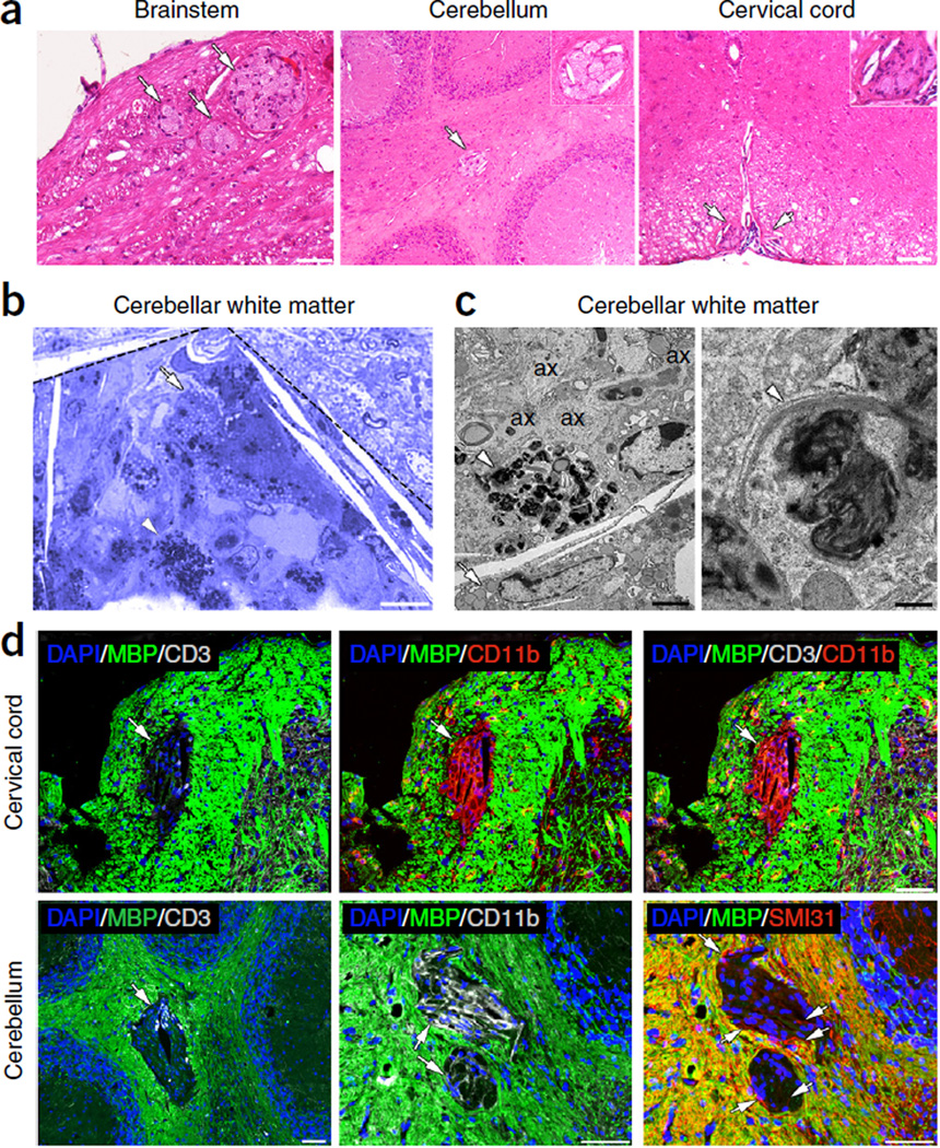
Figure 2.
Focal white matter lesions at early disease stages. (a) Focal white matter lesions (arrows) were detected in different CNS areas of the tamoxifen-treated Plp1-CreERT;ROSA26-eGFP-DTA mice ~40 weeks after injection, such as the brainstem white matter, the cerebellar white matter and the cervical spinal cord white matter. They appear as lighter areas on sections stained with hematoxylin and eosin. Insets show higher magnifications of lesions. Scale bars: 50 µm (brainstem) and 100 µm (cervical cord, cerebellum). (b,c) A focal lesion is outlined in the cerebellar white matter (dashed lines) on a toluidine blue–stained section (b). The lesion contains a high density of macrophages with lipids from degrading myelin (arrow) and myelin debris (arrowhead) in their cytoplasm, which are also shown at higher resolution by EM (c, left). A higher magnification EM image of the myelin debris is shown in c, right. EM analysis also demonstrates the presence of unmyelinated axons (ax) in the focal lesions (c, left panel). Scale bars: 10 µm (b), 2 µm (c, left) and 200 nm (c, right). (d) Focal lesions showed loss of MBP staining in the cerebellar white matter and the cervical spinal cord white matter (green, arrows), and they frequently contained T cells stained for CD3 (gray). These sites also showed increased staining for the microglia and macrophage marker CD11b (red in cervical cord, gray in cerebellum). A few unmyelinated axons were also detected in the white matter lesions by SMI31 staining (red in cerebellum, arrows). Immunofluorescence images are representative of three mice per genotype. Scale bars, 50 µm.

