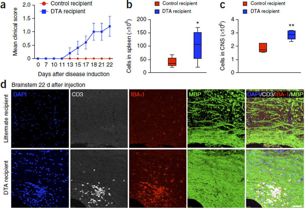
Figure 6.
Adoptive transfer of MOG-specific T cells derived from tamoxifen-treated DTA mice causes white matter inflammation in the CNS of naive Rag1−/− mice. One representative experiment of two is presented. Splenic cells from tamoxifen-treated Plp1-CreERT;ROSA26-eGFP-DTA mice 40–52 weeks after injection or age-matched control (ROSA26-eGFP-DTA) mice were cultured for 72 h at in the presence of MOG35–55 peptide (20 µg/ml) plus IL-12 (10 ng/ml) and IL-2 (100 U/ml). Cultured cells (107 blast cells) from tamoxifen-treated DTA mice were transferred into naive Rag1−/− recipient mice. (a) Mean (± s.e.m.) EAE scores indicated EAE-like neurological symptoms in Rag1−/− mice inoculated with cells from tamoxifen-treated DTA mice (DTA recipient) but not in Rag1−/− mice inoculated with cells from littermate mice (control recipient). (b,c) On day 22 after cell transfer, the number of total cells present in the spleen (b) and CNS (c) were enumerated. (d) Immunohistochemistry revealed the presence of focal inflammatory lesions with high levels of infiltrating T cells (CD3+ cells, gray) and microglia or macrophages (IBA-1+ cells, red), without apparent myelin loss (MBP signal, green) in the brainstem of the Rag1−/− mice inoculated with T cells from tamoxifen-treated DTA mice (bottom). No white matter lesions were detected in those mice inoculated with cells from control littermate (ROSA26-eGFP-DTA) mice (top). Images are representative of five mice per genotype. Scale bar, 60 µm. The data in a–c are presented as mean + s.e.m. One representative experiment of two is presented with n = 5 mice per group; *P < 0.05, **P < 0.01 for differences between control-derived and DTA-derived cell recipient mice shown in b (P = 0.0398) and c (P = 0.0016); one-tailed unpaired Student’s t test.

