
Figure 1.
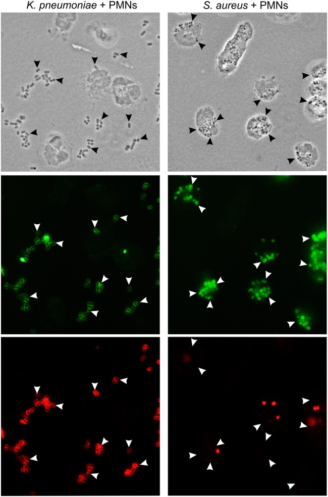
Phagocytosis of multilocus sequence type 258 (ST258) and Staphylococcus aureus. Human neutrophils were combined with Klebsiella pneumoniae strain NJST258_2 (left panels) or S. aureus strain USA300 (right panels), and binding and phagocytosis were evaluated using immunofluorescence microscopy. Top panels are phase-contrast images (arrowheads indicate selected bacteria), middle panels (green staining) show all bacteria, and bottom panels show extracellular bacteria (red staining). Images are representative of the phagocytosis data quantitated in Figure 2. Abbreviation: PMN, polymorphonuclear leukocyte.