
Figure 5.
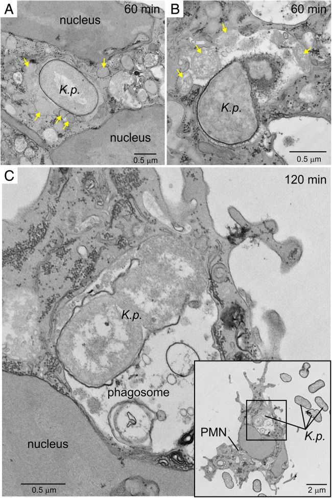
Ultrastructural analysis of ingested Klebsiella pneumoniae. Human neutrophils were cultured with unopsonized K. pneumoniae strain NJST258_2, phagocytosis was synchronized, and samples were analyzed by TEM. A and B, ingested K. pneumoniae 60 minutes after start of the assay. Yellow arrows indicate neutrophil granules, including those that have fused with the K. pneumoniae–containing phagosome. C, Ingested K. pneumoniae 120 minutes after the start of phagocytosis. Destruction of bacteria is evident in panels B and C. Inset, Black square represents the area of the micrograph from which panel C was derived. Abbreviations: K.p., K. pneumoniae; PMN, polymorphonuclear leukocyte.