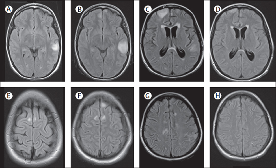
Figure 4. MRI findings in index patient 1.
On day 3 of admission, the MRI of this 16-year-old girl showed multiple cortical-subcortical abnormalities with increased FLAIR and T2 signal involving the left temporal lobe and frontal parasagittal regions (A, E). On day 10, a repeat MRI showed an increase of the size of the temporal lesion and a new cortical lesion in the left frontal lobe (B, F). Repeat MRIs on days 22 and 48 did not show substantial changes (data not shown). Another MRI done 4 months after disease onset showed many new multifocal abnormalities and diffuse atrophy and increase of the size of the ventricles (C, G). A repeat MRI 2 months later, 6 months after symptom onset, showed substantial improvement and resolution of the abnormalities as well as improvement of the ventricular dilatation (D, H).

