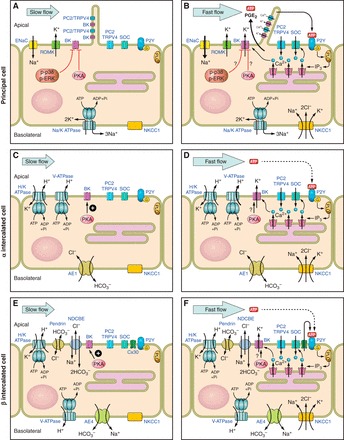
Fig. 2.

Cell-specific mechanoregulation of BK channel-mediated K secretion in the CCD. BK channels, present in principal cell cilia and at the apical membranes of both principal (A and B) and intercalated (C and D, E and F) cells in the CCD, are closed at slow physiologic flow rates (A, C, and E). An increase in tubular flow rate (B, D, and F) induces BK channel-mediated K secretion (FIKS), which requires ENaC-mediated apical Na entry, an increase in intracellular Ca2+ concentration (reflecting internal store release and Ca2+ entry into cells), and basolateral NKCC1 activity. Emerging evidence indicates that BK channel activity in this nephron segment is regulated by autocrine/paracrine factors released into the tubular fluid as well as cell-specific macromolecular complexes that include kinase signaling. Note that we do not know as yet whether principal or intercalated cells mediate FIKS. See manuscript for details.
