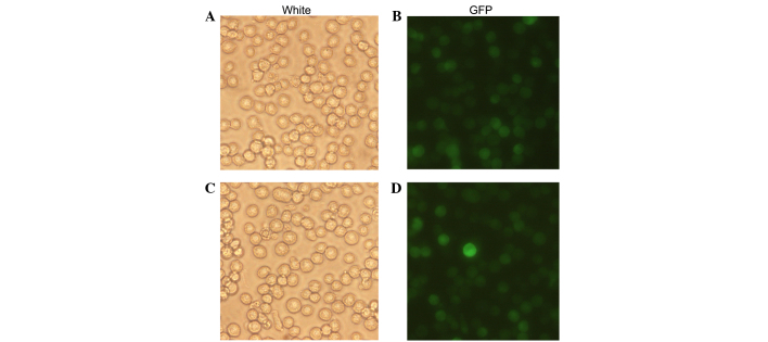
Figure 2.
Fluorescence microscopic assessment of NB4 cell transfection. (A) Light microscopy and (B) fluorescent microscopy images of NB4 cells were all transfected with LV5-NC. (C) Light microscopy and (D) fluorescent microscopy images of NB4 cells were transfected with LV5-NE. Green fluorescent protein expression in infected cells indicated successful expression of lentivirus containing NE. GFP, green fluorescence protein; NE, neutrophil elastase. Magnification, ×200.

